Brain-spine interface
Developing a fully implantable spinal prosthesis

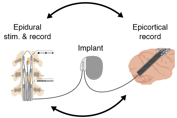
The goal of the fully embedded brain-spinal interface project is to leverage a fully implantable system’s simultaneous recording and stimulation capabilities to develop a single implanted system capable of delivering spatially and temporally precise stimulation to the lumbar spinal cord in order to facilitate stepping. Previous studies in non-human primates have demonstrated that locking stimulation trains to specific phases of the gait cycle restores lost locomotor ability after a spinal-cord injury, and in healthy animals, it increases the step height. They used recordings from implanted microelectrode arrays in the primary motor cortex as the input to a classifier to determine when to apply the stimulation pulses. Here we wanted to replace the intracortical recordings with epidural ECoG, and integrate the recording and stimulation components into a single embedded system with the implanted device.

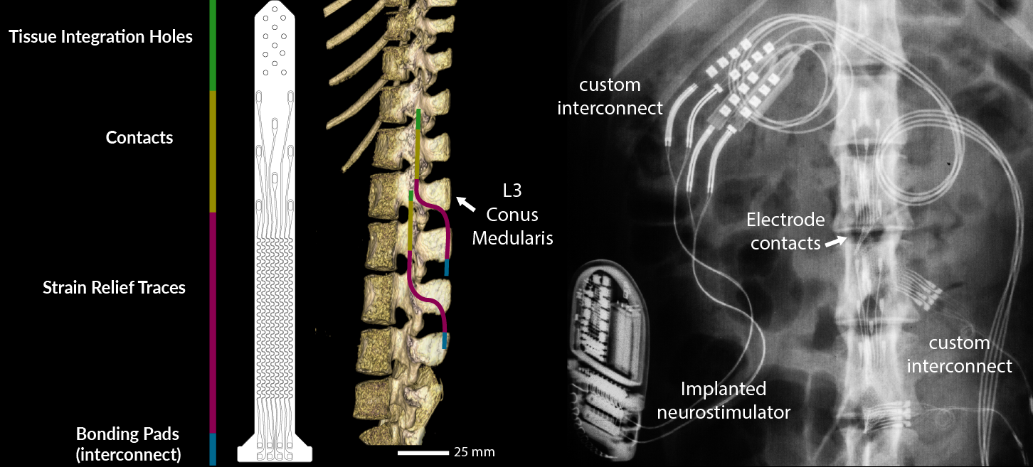
A CAD drawing of the custom electrode paddle used in these experiments. The color-coded line delineates the active (electrode contacts) and inactive (silicone, metallic traces) areas. In the middle, a CT of the animal spine illustrates the surgical plan: two such electrode paddles were implanted to span the area of the lumbar spinal cord where the leg proprioceptive afferents are located. On the right, a post-operative x-ray of the animal with the implanted system.

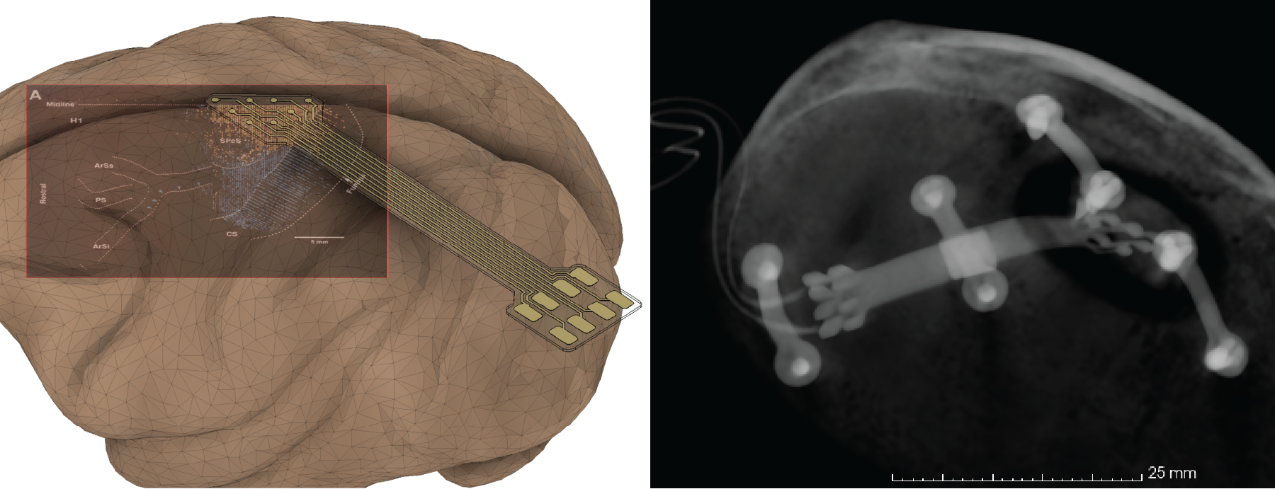
A CAD drawing of the custom electrocorticographic grid I designed for these experiments, superimposed with a reconstructed cortical surface and an illustration of the location of the leg area of motor cortex (He, 1993). On the left, a CT of the device implanted over that site.